
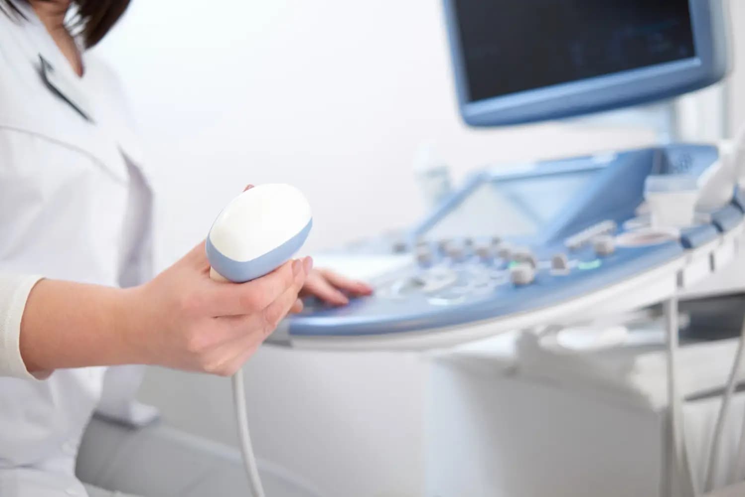

Breast sonography, also known as breast ultrasound, is an imaging examination that helps the doctor assess breast tissue and take a closer look at any changes. The examination uses ultrasound waves and does not involve ionizing radiation.
What has changed since 1 July 2025?
Since 1 July 2025, new rules apply to preventive breast ultrasound examinations. Breast ultrasound is no longer part of the preventive gynecological check-up for women without symptoms, and the preventive examination is not covered by public health insurance.
However, sonography remains an important part of diagnostics. The doctor may recommend it for various changes or difficulties, such as a lump, pain, or changes to the skin or nipple. It is also used to assess certain findings more closely and, if needed, as a complementary examination to mammography.
If you notice a new or unusual change in your breast, it is therefore important to contact your doctor. Depending on the specific situation, they will recommend the appropriate next steps and examinations.
What is a breast ultrasound examination?
Breast sonography (ultrasound) is a simple and painless examination in which the doctor examines the breast tissue using an ultrasound probe. Ultrasound makes it possible to visualize the tissue structure and detect any changes, such as cysts or other lesions.
The examination does not use ionizing radiation. The doctor may recommend it depending on the specific situation – for example, when you have difficulties, a new change in the breast, or as a complement to another examination. The doctor decides whether ultrasound is appropriate based on age, symptoms, findings, and other circumstances.
How to prepare for the examination?
No special preparation is needed for a breast ultrasound. On the day of the examination, you can eat and drink normally.
Before the examination, it is advisable not to apply body lotion, oil, or greasy cream to the breasts and armpits so that the probe can move easily over the skin.
If you have results from previous breast examinations, such as mammography, ultrasound, or biopsy, please bring them with you.
What happens during the examination?
During the examination, the woman lies on her back or slightly on her side with one arm placed above her head. The doctor applies ultrasound gel to the breast and gradually examines the breast tissue with the probe. If needed, the armpit area is checked as well.
The examination is usually painless and takes only a few minutes. The doctor continuously observes the breast tissue and any changes. After the examination, the doctor discusses the result with the patient and, if necessary, recommends the next steps.
When is a breast ultrasound examination appropriate?
Whether a breast ultrasound is necessary depends on the specific situation. The doctor takes into account the woman's age, the character of the breast tissue, any difficulties, the results of previous examinations, and family history.
The doctor may recommend an ultrasound, for example, in case of:
- a new lump or hardening in the breast,
- breast pain or tenderness,
- a change in the shape or appearance of the breast,
- unusual changes to the skin or nipple,
- nipple discharge,
- an unclear finding from another examination,
- the need to assess a change found on mammography more closely.
If you notice a new or unusual change in your breast, it is important to contact your gynecologist or another doctor. Depending on the specific situation, they will recommend the next steps and the necessary examinations.
Ultrasound clinic at Polyclinic Púpava
- MUDr. Katarína Hátašová
More information can be found on the ultrasound clinic page.
Gynecology clinic at Polyclinic Púpava
- MUDr. Jana Štefániková
- MUDr. Olena Mozenbakh
- MUDr. Miriam Elzerová
More information can be found on the gynecology clinic page.
Pay attention to your breasts throughout the year. If you notice a new or unusual change, do not wait for a preventive check-up and consult your gynecologist or doctor.